Home
The preclinical imaging unit offers access to a vast network of experts utilizing modern PET research technology to produce the highest quality results. The unit has a rich history of both industry and academic collaboration over the past 40 years. We have completed countless projects to both our own high standards and our partners’ total satisfaction.
The wide variety of services offered encompass virtually all aspects of PET imaging and we are capable of developing new methods to suit nearly any radiotracer project’s needs. Both academic collaboration and private research contracts are both welcome. We are able to provide services with a wide variety of budgets to suit the means and goals of any partner agency. All stages of research can be offered: from project planning and method development to data analysis and final report writing including everything in between. Examine our expertise page for a more detailed overview of some of the services, technology, and capabilities we have to offer.
The routinely synthesized and available GMP-grade human radiotracer list is vast with countless possibilities for experimental radiotracer syntheses. We operate under strict guidelines with respect to confidentiality to ensure that all information produced for our partners is secure.
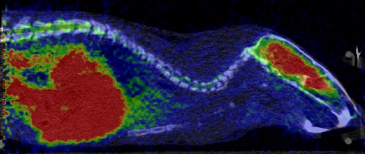
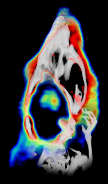
Preclinical imaging is an important step in developing new radiolabelled tracers and a superior tool to study pathophysiology in health and disease. This methodology is powerful for determining many compound characteristics such as receptor occupancy in the early development phases of new pharmaceuticals. Diagnostic values, such as the accuracy, specificity, and sensitivity, of radiotracers can also be evaluated non-invasively and in vivo in animal models.
The advantage of in vivo PET imaging is the time resolution that enables pharmacokinetic properties and tissue biodistribution of the radiolabelled compound to be determined from each animal as a function of time. In longitudinal studies, the development of diseases or the effects of treatments can be efficiently evaluated non-invasively divulging key biochemical changes in the subjects over time.

We regularly work with PET radiotracers labelled with 11C, 18F, 68Ga, and 89Zr and have experience with 15O-labelled tracers. Radiowater studies can be used to acquire perfusion data in target organs and 15O-labelled gases administered via inhalation can be used to determine oxygen metabolism rates in tissues. We have extensive experience with disease models for cancer, neurological disorders, cardiology, inflammation, and metabolic diseases with an enthusiastic and determinate attitude towards new challenges.
The preclinical imaging unit is located in BioCity (www.biocityturku.fi), which is an organization coordinating cutting edge molecular medicine research with state-of-the-art equipment. BioCity core services enable us to combine imaging data with several techniques such as metabolomics, proteomics and single-cell analysis when required. Extensive collaboration with clinicians is a long-lasting tradition and one of our main goal is to efficiently translate preclinical findings to clinical applications. Access to human tissue sections is available via the local biobank (www.auria.fi), which can be utilized to verify in vitro labelled binding assays in human health and disease samples. Several internal core facilities can be accessed to provide expert services and confidential consultation for any project. For example, the professionals at the HistoCore can provide immunohistochemical staining of proteins in tissue sections and the Turku Center for Disease Modeling (www.tcdm.fi) are specialized in producing genetically modified animals. Our animal scanners are located side-by-side to the Central Animal Laboratory (CAL) at the University of Turku. CAL provides services for external clients based on different agreements and, if required, in compliance with OECD guidelines fulfilling GLP (Good Laboratory Practice) requirements.
Feel free to reach out for personalized quotes, budgets, availability and project timelines.


