Technology
STIMULUS project consortium develops new non-invasive, convenient, low-cost, and easy-to-use optical monitoring technology that can measure new digital biomarkers from microcirculation for early detection of cardiovascular diseases. The novel technology will enable earlier detection of heart failure and chronic kidney disease.
For both heart failure and chronic kidney disease patients, it is important to diagnose microvascular dysfunction and monitor the deterioration of microvascular function in early phases of the disease progress. Our proposed technology, the HEMI-speckle system is an integrated solution of two existing prototypes, namely, the multi-spectral optical sensory system and the speckle plethysmography (SPG) system.
Underlying technology I – Multi-spectral optical system for microcirculation hemodynamics, HEMI
A new technique to assess microvascular status has been developed at the University of Turku that is able to measure hemodynamics from large and small arteries to arterioles and capillary bed [1]. This allows the measurement of blood pressure in all tissue layers, vascular resistance, assessment of endothelin function and autoregulation as well as HR, HRV, SpO2 and irregular rhythm.
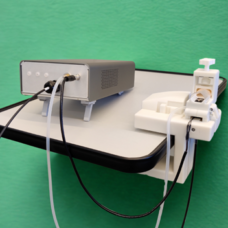
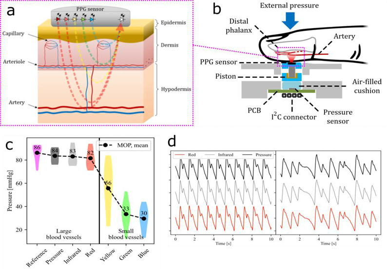
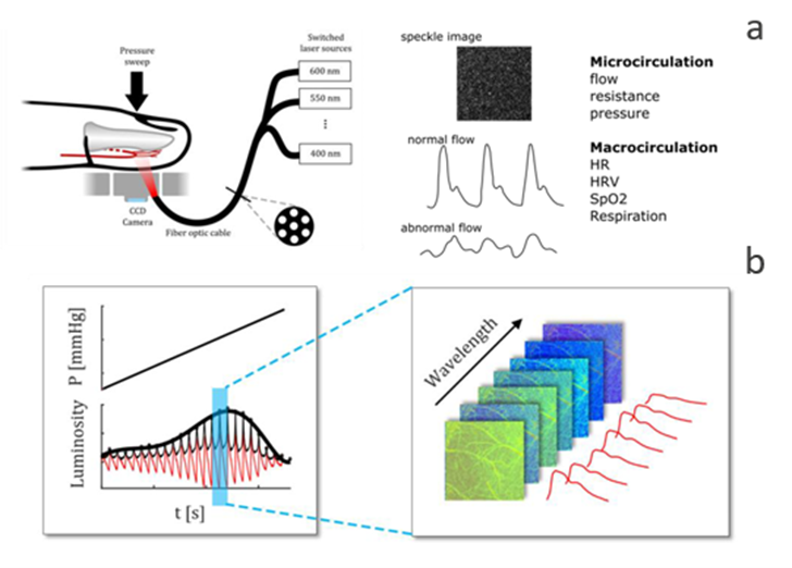
The main operating principle of the HEMI-instrument capable of doing multi-parameter measurements is depicted in the figure below. This is achieved by combining two principles: i) penetration depth of light into tissue is wavelength-dependent, see Figure 1.a, and ii) external compression force exerted on a fingertip alters the underlying vasculature, notably allowing simultaneous measurement of blood pressure at different parts of the vasculature. The developed system comprises a multi-spectral optical sensor, a pressure sensor, and a mechanism to control the external compression force exerted on the fingertip. The measurement is taken by placing a fingertip inside a measurement chamber and then slowly exerting compression force onto it. In the chamber, there is a stacked structure consisting of a printed circuit board with an optical sensor, a piston capable of moving vertically with minimal friction, and an air cushion that couples pressure information to a barometric pressure sensor fixed to a mechanical housing at the bottom of the chamber. The pressing mechanism at the top of the chamber is controlled using a threaded shaft connected to a stepper motor allowing gradual increase of the compression force, see Figure 1.b. Figure 1.c. shows the mean blood pressure for each wavelength and the corresponding arterial or microcirculation hemodynamic each wavelength measures. Figure 1.d. shows signal samples comparing the reference blood pressure with infrared and red hemodynamic measurements.
Underlying technology II – speckle- and photoplethysmography
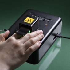
Speckle plethysmography (SPG) is a relatively new technology based on laser speckle contrast imaging. The creation of speckles stems from the use of coherent light (laser light). When coherent light interacts with tissue, random light interaction phenomena are generated leading to the observation of speckles, as depicted in Figure 2.a.

One of the interests of the speckle pattern in the field of biology is when it comes to moving scatterers in tissues, like blood cells for example. These moving scatterers cause the speckle pattern to blur when recorded with an imaging sensor. Using the so-called contrast function and a computational model, we can relate the degree of blurriness to the speed of the scatterers. The calculation of the speckle contrast over a time-series signal leads to the SPG pulse wave, see Figure 2.b.
Photoplethysmography (PPG) is a technique that uses a light source and photodetector and can be used to detect blood volume changes in the microvascular bed of tissue. This is based on the physical property described by the modified Beer-Lambert law which states that the light absorbed by a tissue is proportional to the concentration of fluid present in it. Although PPG is typically obtained using incoherent light (e.g. LED) and a single photodiode, it can also be obtained from the speckle contrast images by calculating the total (or average) intensity. Although the PPG and SPG arise from fundamentally different physical properties of tissue, the proposed technique time-series analysis of speckle contrast imaging provides unique information about both volume and blood flow velocity of the tissue collected by the same photons [2].
Smart toilet
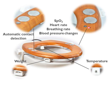
The smart toilet is part of a program called ‘Bathroom for Health’ where sensors are deployed in and around the toilet. This allows for non-obtrusive at home monitoring for preventive health. The toilet seat can already measure several relevant physiological biomarkers, like heart rate and temperature, see Figure 3. Part of this program also explores adding urine analysis inside the toilet. Adding the newly developed technologies to this platform could provide a simple at home tool that can monitor CKD or CVD.
New technology, multi-spectral speckle-based system for microcirculation hemodynamic
The integration of the speckle imaging technique as well as the HEMI will result in a single device, coined as HEMI-Speckle, that can measure a 3D speckle image of the tissue. This innovation is realized by combining the depth resolution technology from multi-spectral optical system with speckle imaging as shown in Figure 4.a. Depth resolution is achieved via multiplexed coherent light sources with different wavelengths. For each coherent wavelength a 2D speckle image is obtained from a different depth of the tissue. The set 2D images are mathematically combined for 3D tissue representation, see Figure 4.b.
References
[1] J-P. Sirkiä, T. Panula, and M. Kaisti “Tonometric Multi-Wavelength Photoplethysmography for Studying the Cutaneous Microvasculature of the Fingertip” IEEE Transactions on Instrumentation and Measurement, vol. 72, (2023)
[2] J. H. Olazabal et al. “Comparison between Speckle Plethysmography and Photoplethysmography during Cold Pressor Test Referenced to Finger Arterial Pressure“ Sensors 24;23(11):5016 (2023)