Left Ventricle (LV)
Obtain a Good PSLAX View
Left ventricular dimensions are measured from the parasternal long-axis view (PSLAX) using either M-mode or a 2D image. M-mode is a standardized measurement technique that allows assessment of cardiac structures and their motion over time. However, M-mode requires good image quality and a favorable cardiac position. If these conditions are not met, measurements should be performed from the 2D image. In patients with significant regional wall motion abnormalities, abnormal left ventricular geometry, or bundle branch block, left ventricular ejection fraction is more reliably assessed by planimetry. For M-mode, the cursor is positioned perpendicular to the long axis of the left ventricle so that it passes just distal to the tips of the open mitral valve leaflets.
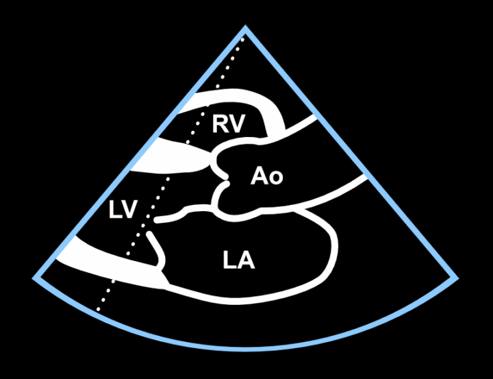
Figure: High-quality PSLAX view and correct M-mode cursor position.
Measurements from the Parasternal View
Once a high-quality PSLAX view and an appropriate M-mode cursor position have been obtained, press the M-mode button on the ultrasound system. Allow the tracing to scroll, and if the recording is of sufficient technical quality, freeze the image using the Freeze button. Measurements are then performed from the frozen image by selecting the desired parameter (e.g. left ventricular internal diameter in diastole) from the measurement menu. On some ultrasound systems this menu is called LV Study. Always measure from tissue to tissue.
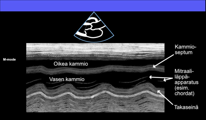
Figure: Basic M-mode view in the PSLAX projection.
RV Measurement in Diastole
The right ventricle (RV) is only rarely measured in this view and projection, but the M-mode measurement is shown here for completeness.
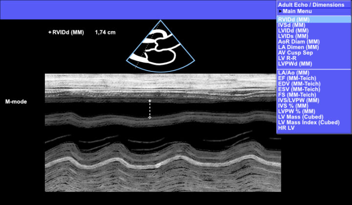
Figure: Right ventricular (RV) measurement using M-mode – a rarely performed measurement.
IVS Measurement in Diastole
Left ventricular measurements are most commonly started by measuring the interventricular septum (IVS).
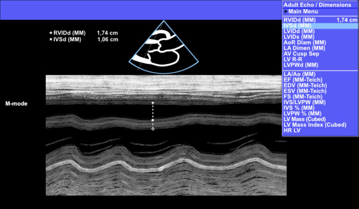
Figure: Interventricular septum (IVS) measurement using M-mode.
LV Measurement in Diastole
Measure LVIDd as shown in the example below.
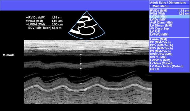
Figure: Left ventricular diastolic diameter (LVIDd) measurement using M-mode.
LVPW Measurement in Diastole
Measure LVPWd as shown in the example below.
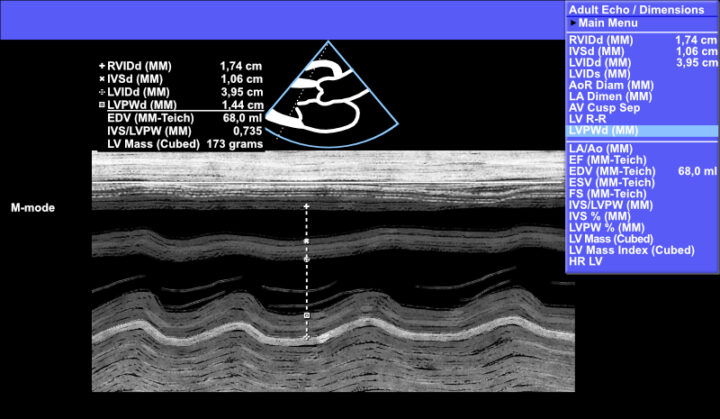
Figure: Left ventricular posterior wall (LVPWd) measurement using M-mode.
LV Measurement in Systole
Measure LVIDs as shown in the example below.
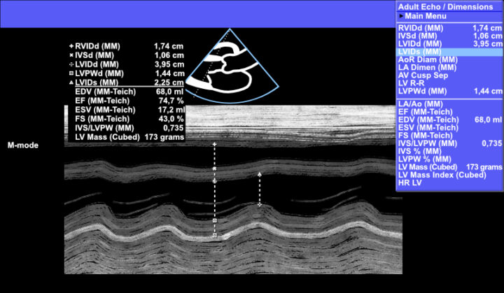
Figure: Left ventricular systolic diameter (LVIDs) measurement using M-mode.
Left Ventricular Measurement Using M-mode
The following video demonstrates how to position the M-mode cursor, slightly increase the gain to improve image definition, and measure IVS, LVIDd, LVIDs, and LVPWd. The ultrasound system then automatically calculates the ejection fraction.
Video: Left ventricular dimensions and function measured using M-mode.
Left Ventricular Measurement Using 2D Imaging
If an adequate M-mode cursor position cannot be obtained, measurements should be performed directly from the 2D image. The most important point is to select LVIDd and LVIDs from Calc → Dimensions, so that the ultrasound system recognizes which measurements are being performed.
Video: Left ventricular measurement using 2D imaging.
Reporting the Results
Once the measurements have been stored by the ultrasound system, it automatically calculates several derived parameters, including EDV, EF, ESV, FS, IVS/LVPW, and LV Mass (Cubed). In the Turku region, it is customary to report only the measured dimensions together with EF. For example, if RV has not been measured, the findings shown below would typically be reported as: “LV 40/23, EF 75%. Walls 11/14.” Dimensions are reported in millimetres without explicitly writing the unit “mm”. Reporting practices vary between hospitals.
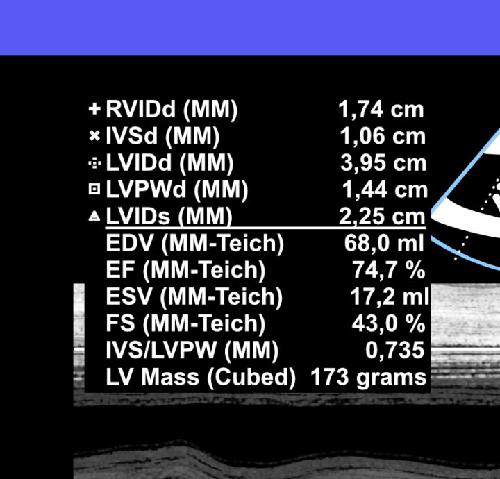
Figure: Left ventricular dimensions and calculated parameters obtained using M-mode.