Apical Extra Views
Alternative Apical Four-Chamber View
If, on the other hand, the transducer marker is directed toward the patient’s body midline, the left ventricle appears on the left side of the image. In this orientation, “the left is on the left and the right is on the right”.
Both imaging orientations for the apical four-chamber view—the standard and the alternative—are acceptable and are used both in our institution and elsewhere according to the echocardiographer’s personal preference. I have personally used this alternative orientation out of habit, which is why several illustrations in this guide are presented from this perspective.
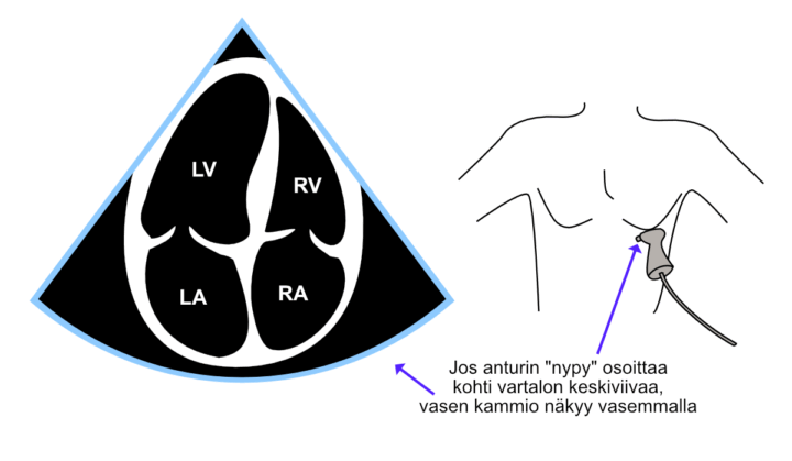
Figure: Apical four-chamber view (A4C) obtained using the alternative orientation.
Apical Views
The heart is commonly examined from three apical views. Comparing these views allows assessment of the left ventricular walls according to the coronary artery territories and helps identify valvular abnormalities that may not be visible in every projection. Starting from the apical four-chamber view, rotating the transducer approximately 60° counterclockwise first produces the apical two-chamber view (A2C), followed by the apical three-chamber view (A3C).
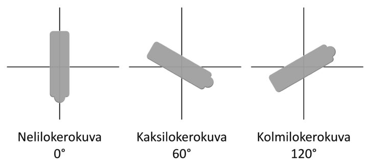
Figure: Apical views obtained by rotating the transducer.
Apical Two-Chamber View (A2C)
The apical two-chamber view is obtained from the apical four-chamber view by keeping the transducer at the cardiac apex and rotating it approximately 60° counterclockwise. This view shows the left ventricle, mitral valve, and left atrium.
Video: Apical two-chamber view (A2C).
Apical Three-Chamber View (A3C)
The apical three-chamber view is obtained from the apical two-chamber view by keeping the transducer at the cardiac apex and rotating it a further 30–60° counterclockwise. This view shows the left ventricle, mitral valve, left atrium, aortic valve, and aortic root.
Video: Apical three-chamber view (A3C).