Inferior Vena Cava (IVC)
Measurement Location
Even a beginner can usually identify the inferior vena cava (IVC) quite easily from the subcostal view. In some patients, however, the subcostal acoustic window does not provide adequate diagnostic image quality, for example because of bowel gas (doi:10.1097/CCE.0000000000001035). If the IVC is not adequately visualized, the patient can be asked to take a deep breath and hold it. This maneuver may lower the diaphragm and improve visualization of the IVC. However, respiratory variation of the IVC cannot be assessed during breath-holding.
I recommend measuring the inferior vena cava (IVC) from the 2D image. In many cases, M-mode does not provide a perfectly perpendicular section of the IVC, and obtaining one often wastes time, especially for beginners. The IVC should not be measured immediately at the hepatic vein junction but approximately 1–3 cm caudal to it, as shown in the figure below. Record the IVC during both expiration and inspiration. There is no need to determine which respiratory phase is being recorded—simply measure the IVC at the same location when it is at its largest and at its smallest.
I also recommend including part of the right atrium in the image whenever possible, since the IVC enters the right atrium, confirming that the correct vessel is being measured. Transducer tilting is also helpful for identifying the IVC.
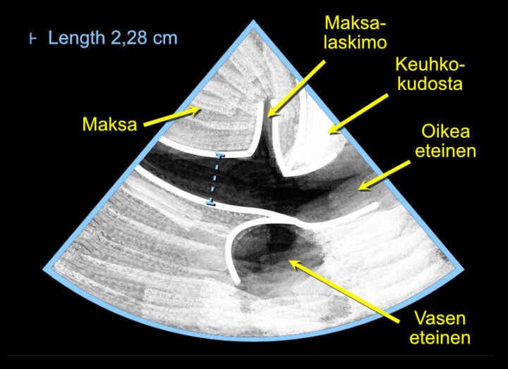
Figure: Measuring the diameter of the inferior vena cava (IVC) during both expiration and inspiration.
Video: Measuring the diameter of the inferior vena cava (IVC) during both expiration and inspiration.
Note! The limitations of IVC measurements for assessing intravascular volume status should be recognized.
I recommend reading, for example, the review by Marik and colleagues on this topic (doi:10.1378/chest.07-2331). Significant tricuspid regurgitation, for example (doi:10.1007/s00134-016-4357-9), may complicate estimation of volume status based on the IVC, something I have also encountered frequently in clinical practice.