Right Ventricle (RV)
Diameter
The right ventricular diameter is measured in diastole immediately distal to the tricuspid annulus (toward the cardiac apex). This basal measurement is abbreviated RVD1. The mid-ventricular diameter, abbreviated RVD2, is encountered less frequently in the literature and even less commonly in routine clinical practice. The measurement locations are shown below.
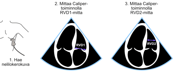
Figure: RVD1 and RVD2 measurements.
Systolic Function
Earlier, left ventricular ejection fraction was estimated using M-mode and 2D measurements. These methods rely on the simplification that the left ventricle contracts predominantly in the radial direction. For the right ventricle, the opposite simplification can be applied: the right ventricle contracts predominantly in the longitudinal direction. Consequently, right ventricular systolic function can be estimated by measuring the longitudinal excursion of the lateral tricuspid annulus. This parameter is known as TAPSE (tricuspid annular plane systolic excursion).
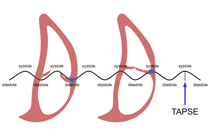
Figure: Definition of TAPSE. When the measurement point (blue dot) is placed on the lateral tricuspid annulus, the movement of the right ventricle between diastole and systole produces a wave-like motion. The amplitude of this motion is TAPSE.
TAPSE Measurement
The measurement is performed as follows:
- Obtain a representative apical four-chamber view in which the M-mode cursor can be aligned from the cardiac apex directly through the lateral tricuspid annulus.
- Activate M-mode.
- The tricuspid annulus produces a wave-like motion. Measure from the peak of the wave to its trough using the Caliper tool. Although the caliper line represents the hypotenuse, the ultrasound system reports the vertical displacement, i.e. the TAPSE value.
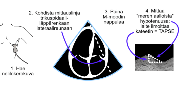
Figure: TAPSE measurement using M-mode. The image shows the alternative apical four-chamber orientation.
TAPSE measurement is generally very easy. The main challenges for beginners are obtaining adequate visualization of the right side of the heart and distinguishing the tricuspid annulus from other right ventricular structures in the M-mode image. Occasionally, a slight counterclockwise rotation of the transducer (approximately 10–20°) improves visualization of the right heart.
Video: TAPSE measurement using M-mode. The video shows the standard apical four-chamber orientation.