Tricuspid regurgitation
Tricuspid regurgitation is usually best assessed from the apical four-chamber view, where the right atrium and right ventricle are visualized in the same imaging plane. The regurgitant jet is first identified using color Doppler, after which its peak velocity is measured with continuous-wave (CW) Doppler. The measurement should always be obtained from the projection in which the Doppler beam is as parallel as possible to the regurgitant jet. For this reason, the optimal view may sometimes be a parasternal or subcostal projection.
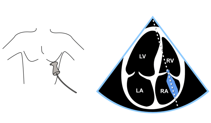
Figure: Assessment of the RV/RA pressure gradient using color Doppler and continuous-wave Doppler from the apical four-chamber view. The image shows an alternative apical four-chamber projection.
Video: Measurement of the RV/RA pressure gradient from the apical four-chamber view. The video shows the standard apical four-chamber projection.
Measuring tricuspid regurgitation is a key echocardiographic parameter, as mild physiological regurgitation is often detectable and can be used to estimate pulmonary artery pressure.