Apical
Apical Four-Chamber View (A4C)
From the apical view—sometimes also called the apex view—a four-chamber view (A4C) is obtained. The standard method is to direct the transducer marker laterally toward the patient’s left side, so that the left ventricle appears on the right side of the image (i.e. “left on the right and right on the left”). This imaging orientation is recommended because it is used internationally and because the automated functions of more advanced ultrasound systems are designed for this orientation (see Additional Apical Views).
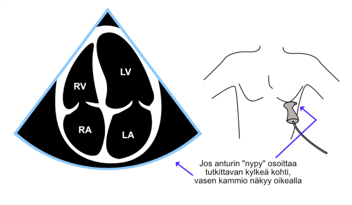
Figure: Apical four-chamber view (A4C) obtained using the standard orientation.
Video: Apical four-chamber view (A4C) obtained using the standard orientation.
Apical Five-Chamber View (A5C)
The apical five-chamber view (A5C) is relatively easy to obtain by first acquiring the four-chamber view and then tilting the transducer slightly ventrally. The four-chamber view is restored by tilting the transducer slightly dorsally.
The main purpose of the five-chamber view is assessment and Doppler measurements of the aortic valve, for example using color Doppler and continuous-wave Doppler.
Sometimes ventral tilting of the transducer causes the acoustic window to disappear completely, or the aorta opens at such an oblique angle that continuous-wave Doppler measurements across the aortic valve cannot be obtained. In these situations, it often helps to slide the entire transducer a short distance along the rib toward the patient’s side (“toward the axilla”).
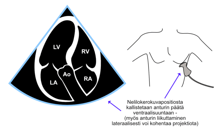
Figure: Apical five-chamber view (A5C) obtained using the alternative orientation (transducer marker toward the midline).
Video: Apical five-chamber view (A5C) obtained using the standard orientation (transducer marker toward the patient’s left side).