Valves
According to the European Society of Cardiology (ESC), aortic stenosis is the most common valvular heart disease requiring an invasive intervention in Europe and North America.
A clinician learning echocardiography soon learns that severe aortic stenosis can often be suspected—or confidently excluded—by visual assessment alone, even before performing quantitative Doppler measurements. In particular, the parasternal long-axis (PSLAX) and parasternal short-axis (PSAX) views are invaluable for assessing leaflet morphology and opening. If the valve appears heavily calcified and leaflet excursion is markedly restricted, aortic stenosis should be suspected.
Color Doppler complements the two-dimensional examination by evaluating blood flow across the valve. When there is no significant obstruction to flow, the color Doppler signal appears uniform, reflecting laminar flow. When blood passes through a narrowed valve orifice, the flow becomes turbulent, producing a characteristic mosaic color Doppler pattern with prominent yellow hues due to aliasing. Whenever turbulent flow is observed across the aortic valve, aortic stenosis should be considered until proven otherwise.
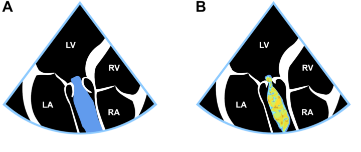
Figure: Laminar (A) and turbulent (B) flow.