Aortic Valve Area (AVA)
- Left ventricular outflow tract (LVOT) diameter
- Blood flow velocity across the aortic valve
- Blood flow velocity in the LVOT
The continuity equation is quite intuitive. We can measure key flow-related parameters at the LVOT, and at the same time we know that blood cannot simply disappear after leaving the LVOT—it must pass through the aortic valve.
Thus, (1) the LVOT diameter (and the automatically calculated LVOT cross-sectional area), together with (2) the amount of blood flowing through the LVOT, allows the echocardiography system to calculate the aortic valve area, since (3) the blood flow across the aortic valve is also known. These measurements are described below.
LVOT Diameter
The first step is to measure the LVOT diameter. The echocardiography system then automatically calculates the LVOT cross-sectional area. The LVOT diameter must be measured structurally from the 2D image.
Note. Do not use the Caliper tool to measure the LVOT diameter, because the echocardiography system will not recognize which structure has been measured.
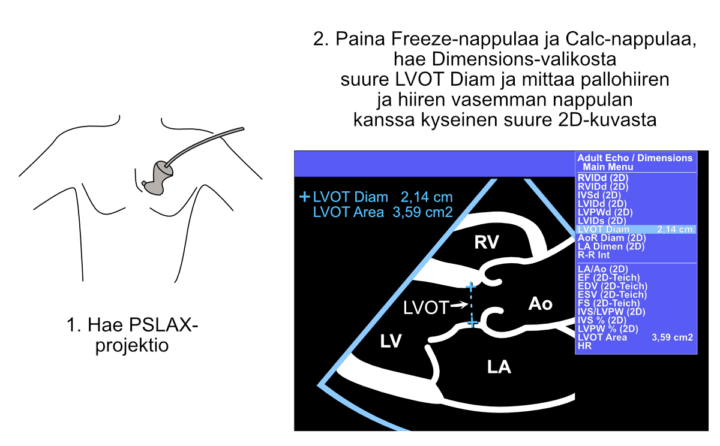
Figure: Measurement of the left ventricular outflow tract (LVOT) diameter.
CW Measurement Across the Aortic Valve
Next, a continuous-wave (CW) Doppler recording is obtained across the aortic valve. Although measuring only the peak velocity would be sufficient for the continuity equation, it is standard practice to trace the entire Doppler envelope. This provides not only the peak gradient but also the mean transvalvular gradient, both of which are routinely reported in echocardiography.
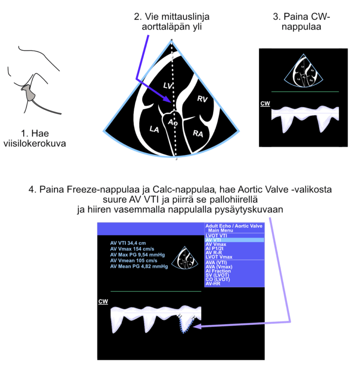
Figure: Continuous-wave Doppler measurement across the aortic valve.
PW Measurement from the LVOT
Finally, blood flow velocity is measured in the LVOT using pulsed-wave (PW) Doppler. Again, although only the peak velocity would be sufficient for the continuity equation, it is standard practice to trace the entire Doppler envelope.
As a useful by-product, the echocardiography system also calculates the stroke volume (SV), which can be included in the echocardiography report. The AVA and SV are displayed automatically once the LVOT diameter, aortic valve Doppler recording, and LVOT Doppler recording have all been measured structurally.
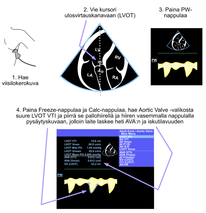
Figure: Pulsed-wave Doppler measurement in the left ventricular outflow tract (LVOT).
If the echocardiography system does not report the stroke volume or AVA, something has gone wrong. Repeat the measurements carefully, making sure to avoid the Caliper tool for all of these measurements.