Subcostal
Inferior Vena Cava (IVC)
The subcostal view is obtained by placing the transducer vertically just caudal to the xiphoid process, with the transducer marker pointing cranially. A slight cranial tilt toward the heart often helps bring the right atrium into view. In addition, a slight tilt toward the liver, which I refer to as the “liver tilt”, may help visualize the inferior vena cava (IVC).
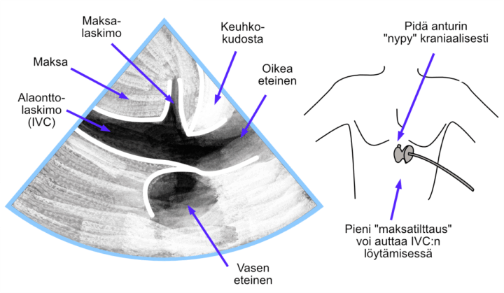
Figure: Subcostal view.
Video: Subcostal view.
Abdominal Aorta
I have noticed that beginners sometimes confuse the inferior vena cava with the abdominal aorta during measurements. To avoid this, I use a simple principle for distinguishing the two:
- First, identify the inferior vena cava using the “liver tilt”.
- Then identify the abdominal aorta using the “spleen tilt”.
Once both the inferior vena cava and the abdominal aorta have been identified, they are easy to distinguish from each other.
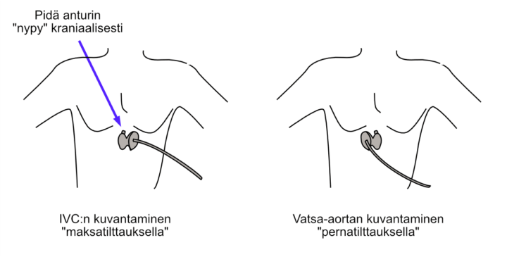
Figure: Identification of the inferior vena cava and abdominal aorta using the “liver tilt” and “spleen tilt”.
Video: Identification of the inferior vena cava and abdominal aorta using the “liver tilt” and “spleen tilt”.