Parasternal
Parasternal Long-Axis View (PSLAX)
The parasternal long-axis view (PSLAX) is obtained by placing the transducer immediately to the left of the mid sternum. The transducer marker should point toward the patient’s sternum / right axilla.
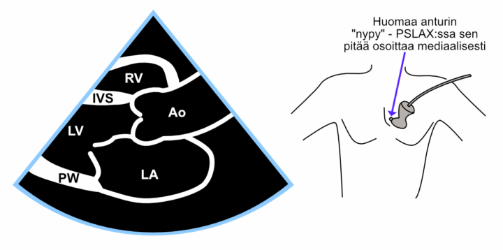
Figure: Parasternal long-axis view (PSLAX).
Video: Parasternal long-axis view (PSLAX).
Parasternal Short-Axis View (PSAX)
The parasternal short-axis view (PSAX) is obtained by rotating the transducer 90° clockwise from the parasternal long-axis view. Sometimes the transducer position needs to be adjusted slightly to restore the acoustic window between the ribs that may have been lost during rotation.
From this view, the entire left ventricle can be scanned step by step by tilting (not rotating!) the transducer either toward the aorta or toward the cardiac apex. At the extreme position toward the aorta, a cross-sectional view of the aortic valve is obtained. At the opposite extreme toward the apex, only the left ventricular apex is seen as a small donut-shaped cross-section.
The image below shows the parasternal short-axis view at the level of the left ventricular papillary muscles. The lower image demonstrates the different tilt positions of the PSAX view (the rotation angle remains constant at 90° clockwise from PSLAX). The video shows the view at four imaging levels, of which the most important are the aortic valve level and the papillary muscle level.
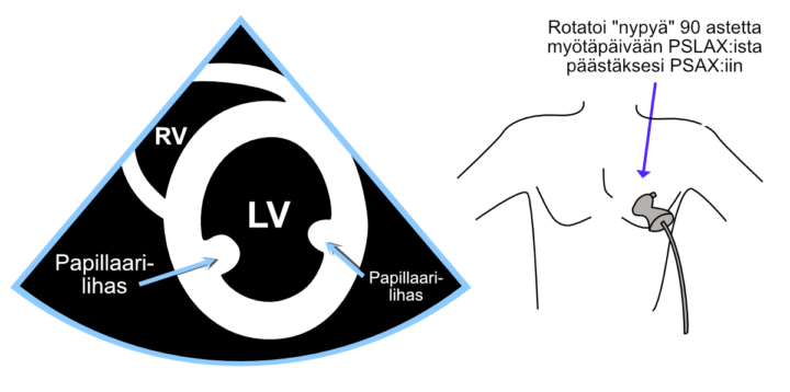
Figure: Parasternal short-axis view (PSAX).
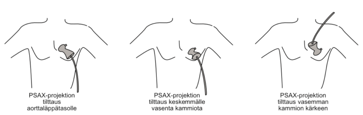
Figure: Tilting the parasternal short-axis view to different imaging levels.
Video: Parasternal short-axis view (PSAX) at four imaging levels.