Lungs
Pleural Effusion
I recommend assessing pleural effusion by placing the transducer vertically on the patient’s mid-axillary line. I prefer to keep the transducer marker pointing toward the axilla, so that the liver appears on the left side of the image and the lung on the right. This orientation makes image interpretation straightforward.
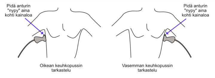
Figure: Left and right scanning positions for pleural assessment.
Lung tissue is easily distinguished from other tissues because it often contains a few comet-tail artifacts (B-lines), which are discussed below. B-lines are not seen in the liver, for example.
If pleural aspiration is being considered, the distance between the lung and the planned puncture site should be assessed using the Caliper tool. I recommend consulting the literature—and, above all, an experienced colleague—regarding safe puncture margins.
Note! If pleural effusion is suspected but cannot be identified with ultrasound, move the transducer as far posteriorly as possible, practically until it touches the examination table or bed sheet. In the supine patient, pleural fluid collects posteriorly under the influence of gravity.
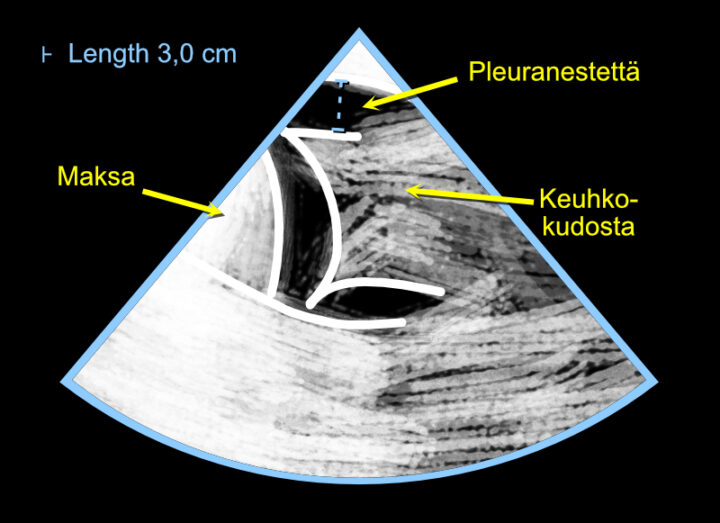
Figure: Assessment of pleural effusion using a cardiac ultrasound system.
Video: Normal pleural space on the right side.
B-lines
The amount of extravascular lung water is assessed from B-lines. These are comet-tail-like vertical artifacts seen on lung ultrasound that can be used in clinical diagnosis. Some characteristic features of B-lines include:
- They appear as vertical comet-tail artifacts rather than horizontal lines.
- They move with the pleural line as the visceral and parietal pleura slide against each other (I like to describe them as being “like windshield wipers”).
- They extend from the pleural line to the bottom of the image without fading.
According to Lichtenstein and Mezière, three or more B-lines in a single scanning view (practically, one intercostal space) is abnormal. Their excellent article, freely available through PubMed Central, provides numerous illustrations and recommended scanning locations.
Via and colleagues have also published an excellent review with many illustrative images, freely available from Minerva Anestesiologica.
These articles also emphasize an important point of differential diagnosis: B-lines are not specific for pulmonary congestion and may also occur, for example, in pneumonia.
B-lines are a good example of Goethe’s observation: “You see only what you know.” Many echocardiographers encounter B-lines thousands of times while performing cardiac ultrasound examinations without recognizing their clinical significance.
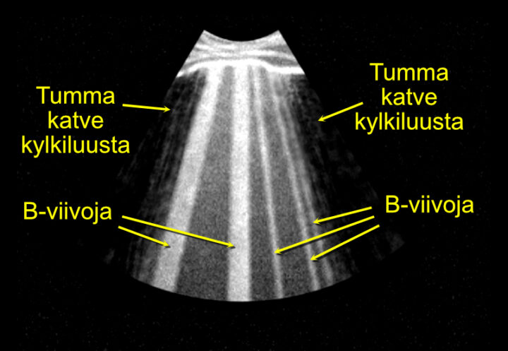
Figure: Assessment of pulmonary congestion by identifying B-lines.
Video: Normal lung ultrasound appearance between the rib shadows. Lung tissue cannot be assessed directly with ultrasound because ultrasound waves do not propagate through air-filled lung. Instead, normal lung ultrasound relies on evaluation of the pleural line and the artifacts arising beneath it.
Lung Sliding
Lung sliding is the physiological movement of the visceral pleura against the parietal pleura during respiration. On ultrasound, this appears as a to-and-fro movement of the pleural line synchronized with breathing. Absence of lung sliding indicates that the lung is no longer expanding normally against the chest wall. The most common cause is pneumothorax, although pleural adhesions, one-lung ventilation, severe atelectasis, or very shallow breathing may also abolish lung sliding. In the appropriate clinical context, absent lung sliding strongly raises the suspicion of pneumothorax.
Video: Normal lung sliding.