Tissue Doppler (TDI) at the Mitral Annulus
When assessing diastolic function, position the sample volume with the trackball immediately lateral to the mitral annulus, in the myocardium between the left atrium and left ventricle. Press the TDI button on the echocardiography system, followed immediately by the PW button. In tissue Doppler mode, the E- and A-waves appear inverted, with downward deflections (Figure 9.3). In this setting they are referred to as the E′-wave and A′-wave (pronounced “E prime” and “A prime”).
Although the E′-wave can also be measured at the septal (medial) mitral annulus, this guide focuses solely on the lateral measurement technique for simplicity.
Note. In patients with atrial fibrillation, there is no organized atrial contraction, and therefore no A′-wave is present.
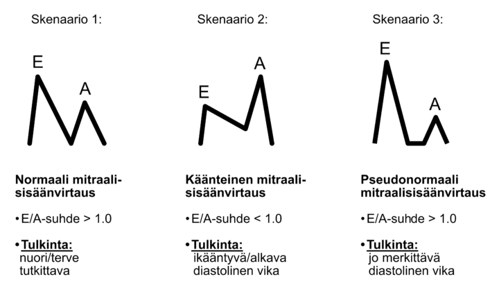
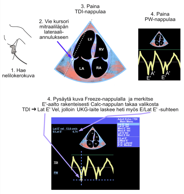
Figure: Tissue Doppler imaging (TDI) for the assessment of diastolic function. The blue-red color map indicates that tissue Doppler mode is active. Because both E′- and A′-waves are present, the patient is in sinus rhythm.
Video: Tissue Doppler imaging (TDI) for the assessment of diastolic function.
TDI Interpretation
When both the E-wave and E′-wave have been measured using the structured Calc workflow, the echocardiography system automatically calculates the E/E′ ratio (often displayed as E/Lat E′ when the lateral annulus is used).
Studies have shown that the E/E′ ratio correlates with pulmonary capillary wedge pressure (PCWP) and therefore serves as a non-invasive estimate of left ventricular filling pressure.
An E/Lat E′ ratio greater than 13 is considered abnormal and suggests elevated left ventricular filling pressure (diastolic dysfunction), according to current ASE/EACVI recommendations.